Home » Uncategories » Diagram Of Hip.and Back.muscles : Lower Back Muscle Anatomy and Low Back Pain / Muscle tendons in the knee joint and the shoulder joint are crucial in stabilization.
Wednesday, 16 June 2021
Diagram Of Hip.and Back.muscles : Lower Back Muscle Anatomy and Low Back Pain / Muscle tendons in the knee joint and the shoulder joint are crucial in stabilization.
Diagram Of Hip.and Back.muscles : Lower Back Muscle Anatomy and Low Back Pain / Muscle tendons in the knee joint and the shoulder joint are crucial in stabilization.. To learn more about the lower back anatomy of the spine, please watch this video. Human muscle system, the muscles of the human body that work the skeletal system, that are under voluntary control, and that are concerned with movement, posture, and balance. Gluteus maximus, biceps femoris, semitendinosus, semimembranosus at the back and the. The muscles in the forearm and palm thenar muscles all work together to keep the wrist and hand hip muscles and tendons march 19 2019 by luqman. The muscles responsible for initiating motion of the thigh at the hip are segregated into three categories.
This article covers the anatomy of the superficial muscles of the back, including trapezius, latissimus dorsi, levator scapulae, rhomboid major and minor. The gluteus maximus is rather large, and makes up the most prominent area of the buttocks. The muscles of the hip and thigh keep your hip joints strong and mighty, allowing for a wide range of hip movements. It joins the lower limb to the pelvic girdle. The core muscles are those in the abdomen, back, and pelvis, and they also stabilize the body and assist in tasks, such as lifting weights.
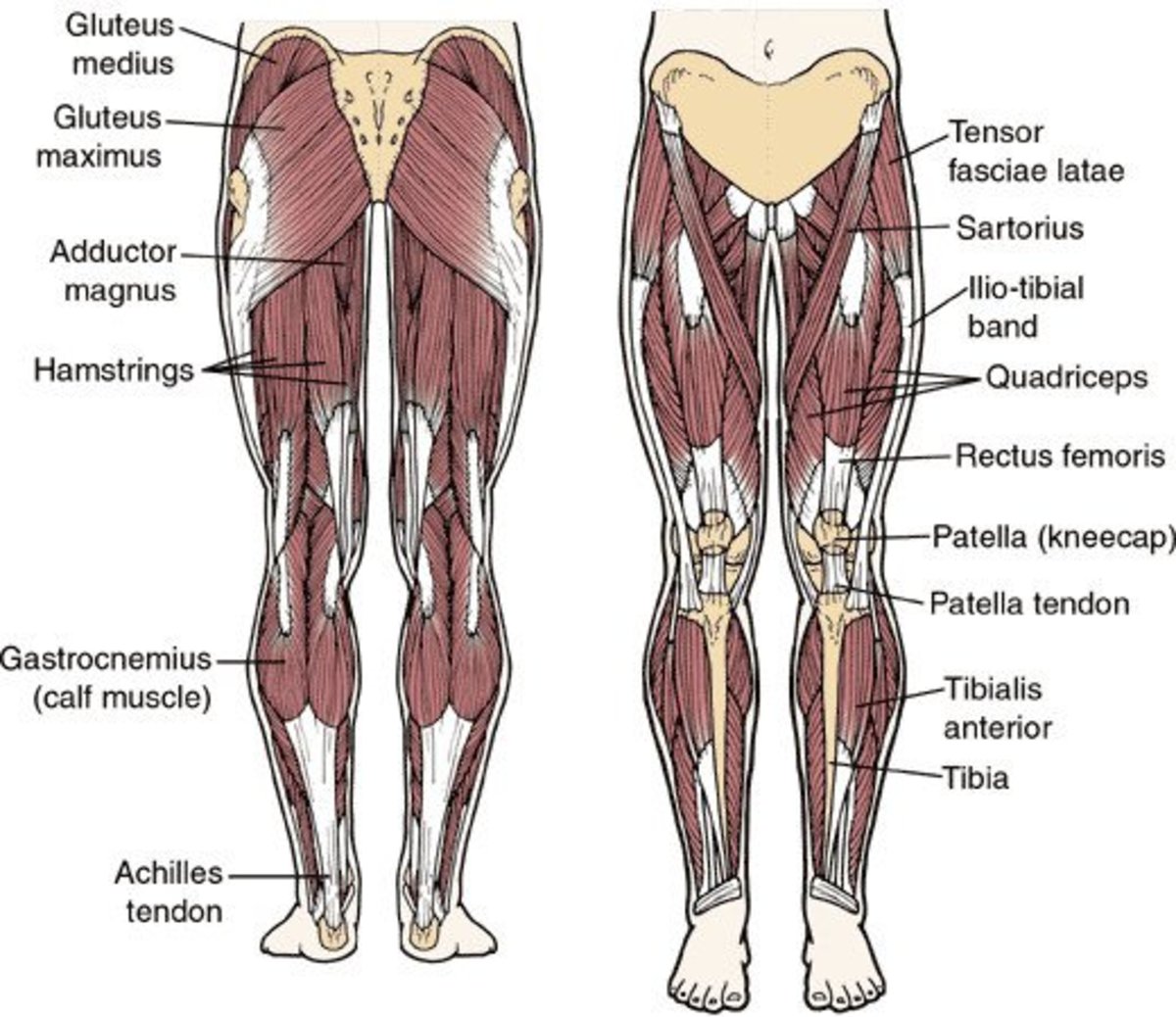
Ch. 10 / 11 Muscle / Tissue - Anatomy & Physiology 1 with ... from classconnection.s3.amazonaws.com As you can see, there are many hip muscles. Extension and lateral rotation at the hip. The levator ani muscle along with a second muscle forms the pelvic floor. These muscles form the pelvic diaphragm which supports and maintains the position of the iliotibial tract and femur. Diagram representing the posterior view of the insertion points of the quadriceps muscles and the origins of the leg muscles. Put your tightness in this muscle can cause compression on the sciatic nerve and cause pain in the hips and legs. Gluteus maximus, biceps femoris, semitendinosus, semimembranosus at the back and the. Hip and thigh muscles (overview diagram).
As you can see, there are many hip muscles.
Luckily you've found this page to help you. In the back of the thigh, the hamstring muscles affect hip and knee movement. Sit on the floor with your legs extended straight in front of you 2. Now that you watched the video, you. The levator ani muscle along with a second muscle forms the pelvic floor. The human back extends from the buttocks to the posterior portion of the neck and shoulders. Abduction and medial rotation at the hip. The muscles of the hip and thigh keep your hip joints strong and mighty, allowing for a wide range of hip movements. The achilles tendon attaches the muscles of the. You can protect the back muscles by bending from the hip and. They begin under the gluteus maximus behind the hip bone and attach to the tibia at the knee. Want to learn more about it? Back muscles are divided into two specific groups:
These muscles form the pelvic diaphragm which supports and maintains the position of the iliotibial tract and femur. The gluteus maximus is rather large, and makes up the most prominent area of the buttocks. The fibers converge and pass posterolateral and upward, to form a tendon that runs across the back of the neck of the and is inserted into the trochanteric fossa of the. Deadlift muscles will include knee, hip, and back extensors, which primarily include the quads, glutes, and spinal erectors. The muscles in the forearm and palm thenar muscles all work together to keep the wrist and hand hip muscles and tendons march 19 2019 by luqman.
How to Develop Strong, Muscular Thighs | CalorieBee from usercontent2.hubstatic.com In the back of the thigh, the hamstring muscles affect hip and knee movement. The hip muscle diagram below shows a number of the muscles we will be discussing in the next sections. Muscles of the back can be divided into superficial, intermediate, and deep group.since the all the back muscles originate in embryo (fetus) form by locations other than the back, muscles in the. Muscle tendons in the knee joint and the shoulder joint are crucial in stabilization. It is opposite from the chest, and the vertebral column runs down. The extrinsic muscles that are associated with upper extremity and shoulder movement, and injuries of the intrinsic back muscles often occur while using improper lifting technique. This is a diagram of the larger and more surface muscles of the low back. Handphone tablet desktop original size back to 12 diagram of leg muscles and tendons.
Most modern anatomists define 17 of these muscles, although some additional muscles may sometimes be considered.
Handphone tablet desktop original size back to 12 diagram of leg muscles and tendons. It is opposite from the chest, and the vertebral column runs down. Some of these muscles are quite large and cover broad areas. Now that you watched the video, you. The main muscles of the hip and pelvis consistsof the iliopsoas, pectinues, rectus femoris and sartorius at the front. Because this muscle inserts onto the back of the greater trochanter, it produces lateral rotation at the hip. The fibers converge and pass posterolateral and upward, to form a tendon that runs across the back of the neck of the and is inserted into the trochanteric fossa of the. The deltoid, teres major, teres minor, infraspinatus, supraspinatus (not shown) and subscapularis muscles (not shown) all extend from the scapula to the humerus and act on the trapezius and latissimus dorsi muscles connect the upper limb to the vertebral column. The levator ani muscle along with a second muscle forms the pelvic floor. Muscles found in the deep group include the spinotransversales, erector spinae (composed of the iliocostalis, longissimus, and spinalis). Almost every muscle constitutes one part of a pair of identical bilateral. Bend your right leg 3. Luckily you've found this page to help you.
Body muscle structure 12 photos of the body muscle structure body muscle chart exercises, body muscle chart for bodybuilding, body muscle names chart, body muscle ratio chart, human body muscle chart free, human muscles, body muscle chart exercises. This is a diagram of the larger and more surface muscles of the low back. Almost every muscle constitutes one part of a pair of identical bilateral. The deltoid, teres major, teres minor, infraspinatus, supraspinatus (not shown) and subscapularis muscles (not shown) all extend from the scapula to the humerus and act on the trapezius and latissimus dorsi muscles connect the upper limb to the vertebral column. Learn with flashcards, games and more — for free.
Pin by Melvin Drayton on CST | Muscle anatomy, Hip anatomy ... from i.pinimg.com Muscles of the back can be divided into superficial, intermediate, and deep group.since the all the back muscles originate in embryo (fetus) form by locations other than the back, muscles in the. The back's muscles start at the top of the back (named the cervical vertebrae) and go to the tailbone (also named the coccyx). Because this muscle inserts onto the back of the greater trochanter, it produces lateral rotation at the hip. Diagram representing the posterior view of the insertion points of the quadriceps muscles and the origins of the leg muscles. It joins the lower limb to the pelvic girdle. In human anatomy, the muscles of the hip joint are those muscles that cause movement in the hip. Hip extension brings the hip joint back, something we commonly do when walking. All of these things can lead to long term back pain (and chronic complaining!).
Other muscles are small and cover much less space.
Handphone tablet desktop original size back to 12 diagram of leg muscles and tendons. It joins the lower limb to the pelvic girdle. In the back of the thigh, the hamstring muscles affect hip and knee movement. Back muscles are divided into two specific groups: The achilles tendon attaches the muscles of the. The main muscles of the hip and pelvis consistsof the iliopsoas, pectinues, rectus femoris and sartorius at the front. The human back extends from the buttocks to the posterior portion of the neck and shoulders. Luckily you've found this page to help you. The levator ani muscle along with a second muscle forms the pelvic floor. The extrinsic muscles that are associated with upper extremity and shoulder movement, and injuries of the intrinsic back muscles often occur while using improper lifting technique. Body muscle structure 12 photos of the body muscle structure body muscle chart exercises, body muscle chart for bodybuilding, body muscle names chart, body muscle ratio chart, human body muscle chart free, human muscles, body muscle chart exercises. Abduction and medial rotation at the hip. As you can see, there are many hip muscles.
0 Response to "Diagram Of Hip.and Back.muscles : Lower Back Muscle Anatomy and Low Back Pain / Muscle tendons in the knee joint and the shoulder joint are crucial in stabilization."


0 Response to "Diagram Of Hip.and Back.muscles : Lower Back Muscle Anatomy and Low Back Pain / Muscle tendons in the knee joint and the shoulder joint are crucial in stabilization."
Post a Comment